3. 展望
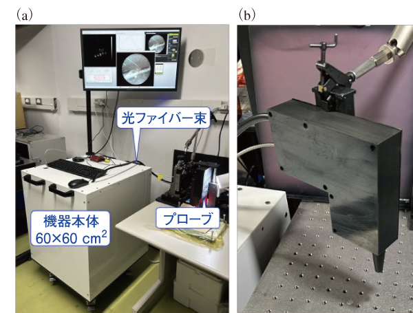
前節において紹介した機械学習モデルを最適化することにより,末梢神経とその他3種類の組織とを正確度95%で判別できる可能性を見出している。このように末梢神経を非侵襲的に無標識で正確に鑑別できる技術は,術中の神経温存において役立つと筆者は考えている。低侵襲化が進む昨今の外科手術は,病変を残さず摘出しつつ神経を可能な限り温存し,術後の患者の良好な予後と高いクオリティー・オブ・ライフを両立しようとしている。しかし,筆者が多数の臨床医らに対し行ったヒアリングによって,一部の手術手技・症例では,細い線維状組織を神経か否か鑑別することは容易ではないことがわかってきた。また,神経の損傷を原因と疑う術後合併症の発生が報告されている14〜17)。関心領域選択的ラマン分光分析技術を応用する医療用末梢神経検知デバイスを実現することにより,術中の神経損傷を原因と疑う術後機能障害の発生を抑止できるようになると筆者は考えている。この考えのもと,泌尿器外科医らとともに,測定プローブを有する末梢神経温存ナビゲーション機器のプロトタイピングに挑戦している(図5)。ラマン分光法を応用した医療機器は,世界をみても成功例は少なく,筆者らの挑戦は容易ではない。実際に,プローブの小型化などの技術的な点だけではなく,機器のコスト面などの事業化に関する点においても課題は山積している。ラマン分光法を応用する未来の医療の実現に向けて,本稿を読まれた皆様にも筆者らの取り組みを応援していただけると幸いである。
謝辞
本稿において紹介した研究の一部は,日本医療研究開発機構(AMED)の医療機器等研究成果展開事業(22hma922004,23hma322013,24hma322013),橋渡し支援プログラム(22ym0126815, 23ym0126815),テルモ生命科学振興財団の研究開発助成(開発助成),及び日本学術振興会(JST)の共創の場形成支援プログラム(COI-NEXT)(JPMJPF2009)の支援のもと実施されたものである。また,本稿において紹介した研究の一部は,京都府立医科大学の髙松哲郎先生,田中秀央先生,浮村理先生,原田義規先生,藤原敦子先生,大阪大学の藤田克昌先生,渡利彰浩先生,冨岡穣先生,南川丈夫先生,坂田渉氏,耿君安氏,サイエンスエッジ㈱の太田泰輔代表取締役らの協力により実施されたものである。この場において心からの感謝の意を表する。
参考文献
1)Z. Huang, S. K. Teh, W. Zheng, J. Mo, K. Lin, X. Shao, K. Y. Ho, M. Teh, K. G. Yeoh, Integrated Raman spectroscopy and trimodal wide-field imaging techniques for real-time in vivo tissue Raman measurements at endoscopy, Opt. Lett. 34, 758-760 (2009).
2)K. Hirose, T. Aoki, T. Furukawa, S. Fukushima, H. Niioka, S. Deguchi, M. Hashimoto, Coherent anti-Stokes Raman scattering rigid endoscope toward robot-assisted surgery, Biomed. Opt. Express 9, 387-396 (2018).
3)C. Shu, W. Zheng, Z. Wang, C. Yu, Z. Huang, Development and characterization of a disposable submillimeter fiber optic Raman needle probe for enhancing real-time in vivo deep tissue and biofluids Raman measurements, Opt. Lett. 46, 5197-5200 (2021).
4)B. Qiu, C. Shu, Z. Huang, Development of a multi-needle fiberoptic Raman spectroscopy technique for simultaneous multi-site deep tissue Raman measurements in the brain, Opt. Lett. 48, 4396-4399 (2023).
5)R. Zhang, R. Bi, C. H. J. Hui, P. Rajarahm, U.S. Dinish, M. Olivo, A portable ultrawideband confocal Raman spectroscopy system with a handheld probe for skin studies, ACS Sens. 6, 2960-2966 (2021).
6)Endofotonics Pte Ltd – Product: m.endofotonics.com/col.jsp?id=104 (25/Jun/2024).
7)RiverD International B.V. – Skin Analysis – Products: www.riverd.com/skin-analysis-with-the-gen2-sca/products/gen2-sca/ (25/Jun/2024).
8)F. Daoust, T. Nguyen, P. Orsini, J. Bismuth, M.-M. de Denus-Baillargeon, I. Veilleux, A. Wetter, P. Mckoy, I. Dicaire, M. Massabki, K. Petrecca, F. Leblond, Handheld macroscopic Raman spectroscopy imaging instrument for machine-learning-based molecular tissue margins characterization, J. Biomed. Opt. 26, 022911 (2021).
9)Y. Kumamoto, M. Li, K. Koike, K. Fujita, Slit-scanning Raman microscopy: Instrumentation and applications for molecular imaging of cell and tissue, J. Appl. Phys. 132, 171101 (2022).
10)Y. Kumamoto, Y. Harada, H. Tanaka, T. Takamatsu, Rapid and accurate peripheral nerve imaging by multipoint Raman spectroscopy, Sci. Rep. 7, 845 (2017).
11)L. Jayes, A. P. Hard, C. Séné, S. F. Parker, U. A. Jayasooriya, Vibrational spectroscopic analysis of silicones: A Fourier transform-Raman and inelastic neutron scattering investigation, Anal. Chem. 75, 742-746 (2003).
12)P.A. Bentley, P.J. Hendra, Polarised FT Raman studies of an ultra-high modulus polyethylene rod, Spectrochim. Acta A 51, 2125-2131 (1995).
13)T. Minamikawa, Y. Harada, T. Takamatsu, Ex vivo peripheral nerve detection of rats by spontaneous Raman spectroscopy, Sci. Rep. 5, 17165 (2015)
14)T. Nakamura, M. Oishi, T. Ueda, A. Fujihara, H. Nakanishi, K. Kamoi, Y. Naya, F. Hongo, K. Okihara, T. Miki, Clinical outcomes and histological findings of patients with advanced metastatic germ cell tumors undergoing post-chemotherapy resection of retroperitoneal lymph nodes and residual extraretroperitoneal masses, Int. J. Urol. 22, 663-668 (2015)
15)I. Schauer, E. Keller, A. Müller, S. Madersbacher, Have rates of erectile dysfunction improved within the past 17 years after radical prostatectomy? A systematic analysis of the control arms of prospective randomized trials on penile rehabilitation, Andrology 3, 661-665 (2015).
16)M. Novackova, Z. Pastor, R. Chmel Jr, T. Brtnicky, R. Chmel, Urinary tract morbidity after nerve-sparing radical hysterectomy in women with cervical cancer, Int. Urogynecol. J. 31, 981-987 (2020).
17)R. Sun, Z. Dai, Y. Zhang, J. Lu, Y. Zhang, Y. Xiao, The incidence and risk factors of low anterior resection syndrome (LARS) after sphincter-preserving surgery of rectal cancer: a systematic review and meta-analysis, Support. Care Cancer 29, 7249-7258 (2021).
■Region-of-interest selective Raman spectroscopy for rapid molecular analysis
■Yasuaki Kumamoto
■Life and Medical Photonics Division, Institute for Open and Transdisciplinary Research Initiatives, Osaka University


